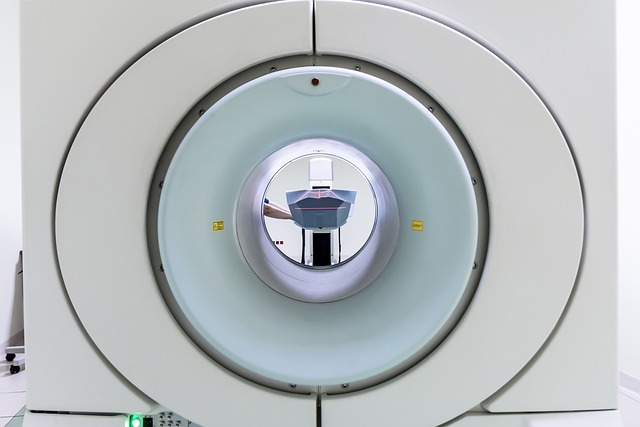
MRI scans utilize magnetic fields and radio waves to create detailed cross-sectional images of the body, with contrast media like gadolinium dyes playing a crucial role in enhancing visualization of soft tissues. These substances optimize signal-to-noise ratios, making it easier to distinguish organs, blood vessels, and soft tissue structures, thus improving diagnostic accuracy and clinical decision-making. Proper administration and selection of contrast media are essential for safe and effective MRI scan contrast use.
“Discover how contrast media enhances the visualization of soft tissues in MRI scans. This comprehensive guide delves into the science behind these substances, their role in improving image quality, and the benefits they offer in diagnosing various conditions. From understanding soft tissues to exploring different types of contrast media and their effects, this article provides insights for both medical professionals and those interested in the power of MRI technology. Learn about the considerations and advantages of using contrast media in medical imaging.”
Understanding MRI Scans and Soft Tissues
Magnetic Resonance Imaging (MRI) scans are a powerful tool in medical imaging, offering detailed insights into the human body’s internal structures. One of its key strengths lies in visualizing soft tissues, which are often challenging to examine using other imaging modalities. Soft tissues, including muscles, organs, and connective tissues, lack distinct structural features visible on traditional X-rays or CT scans.
MRI scans utilize strong magnetic fields and radio waves to generate detailed cross-sectional images of the body. When contrast media, such as gadolinium dyes, are administered, they enhance the signal from specific tissue types, allowing for better differentiation. This process improves the contrast between different soft tissues, making them easier to identify and analyze. As a result, MRI scans provide unprecedented detail in examining organs, blood vessels, and soft tissue structures, leading to more accurate diagnoses and treatment planning.
The Role of Contrast Media in MRI
Contrast media play a pivotal role in enhancing the visualization of soft tissues during Magnetic Resonance Imaging (MRI) scans. These substances are carefully selected to improve the signal-to-noise ratio, enabling clearer and more detailed images of internal body structures. When introduced into the body, contrast media interact with magnetic fields, causing differences in signal intensity that highlight specific tissues on the MRI scan.
This technique is particularly valuable for differentiating between various soft tissue types, such as muscles, fat, and organs, which can be challenging to distinguish using standard MRI protocols alone. By enhancing specific structures, contrast media facilitate a more accurate diagnosis and better understanding of soft tissue abnormalities, thereby improving patient care and clinical decision-making.
How Contrast Enhances Soft Tissue Visualization
Contrast media play a pivotal role in enhancing the visibility of soft tissues during MRI scans, allowing radiologists to gain clearer insights into their structure and function. When introduced into the body, these substances create a distinct difference in signal intensity between the tissues they outline and the surrounding areas, providing high-contrast images. This is particularly beneficial for soft tissues, which can be challenging to discern due to their similar signal properties to surrounding structures like fat or water.
The mechanism behind this enhancement lies in the interaction of the contrast media with magnetic fields. Different types of contrast agents have varying relaxation times, affecting how quickly they release signals after being excited by the MRI machine’s radiofrequency pulses. This variation leads to marked differences in image contrast, making it easier to identify and differentiate soft tissues based on their unique signal characteristics.
Benefits and Considerations of Using Contrast Media
Contrast media play a pivotal role in enhancing the visualization of soft tissues during medical imaging procedures, particularly in magnetic resonance imaging (MRI) scans. One of the primary benefits is improved signal-to-noise ratio, allowing radiologists to better discern subtle details within soft tissue structures. This is especially crucial for detecting abnormalities that might be obscured without contrast agents. For instance, in brain imaging, contrast media can highlight cerebral vascular malformations or tumors that encase surrounding tissues, providing vital information for diagnosis and treatment planning.
However, the use of MRI scan contrast should be carefully considered. Timing and choice of contrast agent are critical; different agents have varying kinetic properties and clearance rates. Inappropriate selection could lead to suboptimal imaging results or even adverse reactions. Additionally, individual patient factors, such as renal function, must be taken into account to ensure safe and effective administration. Proper monitoring during the procedure is essential to optimize image quality while minimizing risks associated with contrast media use.
Contrast media play a pivotal role in enhancing the visualization of soft tissues during MRI scans, providing clearer images that aid in accurate diagnoses. By improving the distinction between various soft tissue types, these agents enable radiologists to detect subtle abnormalities that might be missed without their use. While safe and effective for many applications, the choice and administration of contrast media require careful consideration to balance benefits against potential risks. Ongoing research continues to refine contrast agent technology, promising even better image quality and patient safety in the future.
