MRI contrast injection is a vital tool for neurologists, enhancing brain and spinal cord visibility to diagnose conditions like tumors and demyelinating diseases. Contrast agents interact with magnetic fields to highlight structural differences, revealing intricate details not visible on standard scans. This technique aids in accurate treatment planning for complex neurological disorders while ensuring patient safety through rigorous testing and monitoring.
Contrast agents play a pivotal role in enhancing the visibility and differentiation of brain and spinal cord structures during magnetic resonance imaging (MRI). This technology is crucial for accurate diagnosis, as it enables radiologists to distinguish between various tissues. In this article, we explore why contrast is essential, focusing on ‘Enhancing Tissue Visibility’, ‘Differentiating Structures’, and the unique requirements of brain and spinal cord imaging. Additionally, we delve into the safety aspects of MRI contrast injection.
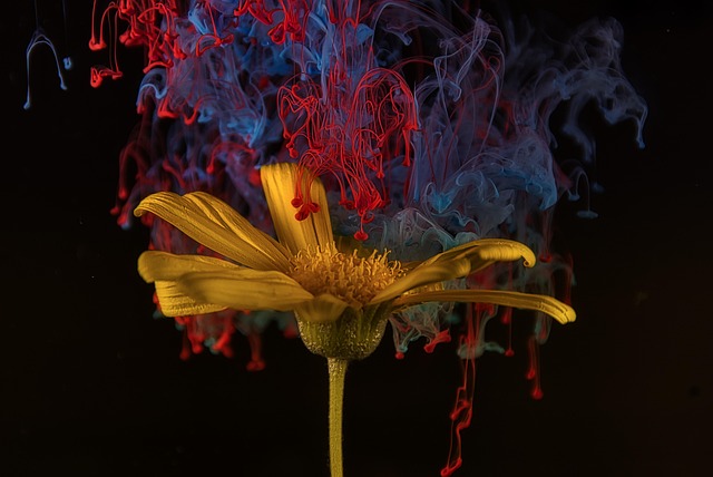
Enhancing Tissue Visibility: The Role of Contrast
Contrast agents play a pivotal role in enhancing tissue visibility during brain and spinal cord imaging via techniques such as MRI. By strategically injecting these agents into the bloodstream, healthcare professionals can highlight specific structures within the complex neural network. This is particularly crucial for differentiating between various types of tissues—gray matter, white matter, cerebral fluid—which appear similar on unenhanced scans.
The mechanism behind this enhancement involves the unique interaction between the contrast agent and magnetic fields in an MRI machine. Contrasting agents, often based on gadolinium or iron compounds, alter the signal intensity detected by the scanner, making certain tissues stand out more clearly. This improved visibility allows radiologists to accurately diagnose conditions like tumors, lesions, or demyelinating diseases—issues that might otherwise be subtle or difficult to discern in standard scans. Moreover, the administration of MRI contrast injection can provide crucial insights into blood flow patterns and vessel integrity, further enriching the diagnostic landscape for neurological disorders.
Differentiating Structures: Key to Accurate Diagnosis
Differentiating structures within the brain and spinal cord is a critical aspect of accurate diagnosis in neurology and neurosurgery. Magnetic Resonance Imaging (MRI) with contrast injection plays a pivotal role here, enabling radiologists to distinguish between various tissues, blood vessels, and pathologies. By enhancing specific structures with contrasting agents, MRI can reveal intricate details that might be imperceptible on standard scans. This is particularly important in identifying brain tumors, cerebral hemorrhages, or demyelinating diseases, where subtle differences in tissue appearance can significantly impact treatment planning.
The choice of contrast agent and its administration protocol are tailored to the specific imaging goal. For example, gadolinium-based agents are commonly used to highlight vascular structures, aiding in the detection of brain lesions or evaluating blood flow. On the other hand, certain agents like manganese-enhanced MRI can improve visualization of gray matter, helping in the assessment of neurological disorders affecting cognitive functions. Effective use of contrast injection, therefore, enhances the diagnostic accuracy and ultimately guides clinicians in making informed decisions for patient care.
Brain and Spinal Cord: Unique Requirements for Imaging
The brain and spinal cord, integral components of the nervous system, pose unique challenges for imaging due to their complex structures and sensitive nature. Unlike other organs, these neural structures require a high level of detail and precision in visualization to accurately diagnose conditions like tumors, lesions, or injuries. This is where MRI contrast injection plays a pivotal role.
Contrasting agents enhance specific aspects of the brain and spinal cord’s appearance on MRI scans, enabling radiologists to differentiate between various tissues and abnormalities. For example, certain contrasts can highlight blood flow, helping to identify vascular malformations, while others may demonstrate changes in tissue density, aiding in detecting tumors or neurological disorders. This tailored approach ensures that even subtle variations within these delicate structures can be captured, providing essential insights for accurate diagnosis and treatment planning.
MRI Contrast Injection: Safety and Considerations
MRI contrast injections play a crucial role in enhancing brain and spinal cord visibility during imaging, but safety is paramount. These specialized agents are designed to temporarily improve the signal-to-noise ratio, allowing radiologists to detect subtle abnormalities that might be obscured by normal tissue. However, as with any medical procedure involving foreign substances, there are considerations to keep in mind.
The safety of MRI contrast injections relies on rigorous testing and regulatory oversight. While rare, potential side effects include allergic reactions, kidney damage due to the contrast agent’s breakdown products, and, in some cases, neurological issues. Patients with certain medical conditions like kidney disease or allergies should be carefully evaluated before administration. Healthcare providers rigorously monitor patients during and after the procedure, ensuring prompt intervention if any adverse reactions occur.
Contrast agents play a pivotal role in enhancing the visibility of brain and spinal cord tissues, enabling accurate diagnosis through magnetic resonance imaging (MRI). By differentiating crucial structures within these complex regions, MRI contrast injection becomes an indispensable tool for healthcare professionals. Safety considerations are paramount, but the benefits far outweigh the risks, making it a game-changer in navigating the intricate landscapes of the brain and spinal cord.